**OFFICIAL GEEK ALERT: if you do not want to know what an “aquaporin” is or what the “corneo-epidermal” junction is, then look away now”
I frequently find that in talking about skincare, we gloss over how the skin itself keeps itself hydrated. It is essential to understand that the skin is a self-regulating mechanism and at (say) 25 years of age, it is operating optimally. However, as we age, our self-regulating mechanism, starts to slowdown. The function of a healthy lifestyle (diet, exercise, UV protection) as well as skincare, is to help the skin restore itself to its optimal modus operandi.
For example, desquamation is a process by which dead skin cells leave the skin and this happens in (e.g.) someone who is 25, every 28 days. But as you get older, this can increase to about 40 days. The problem with accumulating dead skin cells, is that the top layer of the skin becomes drier and actually increases the level of Trans Epidermal Water Loss. In addition, the top layer of our skin forms dry, scaly grids.
A well-organised Stratum Corneum…
The skin has three layers: the epidermis, dermis and hypodermis. The Stratum Corneum (“SC”) (or the skin barrier or the horny layer) is the topmost layer of the epidermis (and the skin). It is what is visible. “Soft and supple” skin has tightly knit corneocytes organised in a flattened manner.
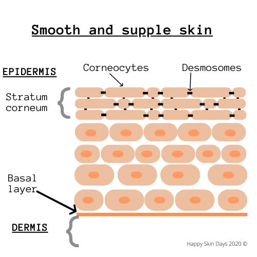
This alignment of the corneocytes in the SC is what makes skin “soft” and “supple.” The NMF and lipids are at peace (or homeostasis) with the other elements around them. Both are essential for a proper functioning skin barrier. One of these crucial functions is reducing the amount of TEWL from the epidermis and keeping the skin hydrated.
If there were no Stratum Corneum, then we would dry out.
If the skin is dry (e.g., low humidity, age, medical condition such as diabetes etc.), then the corneocytes are not aligned tightly.
Today’s blog
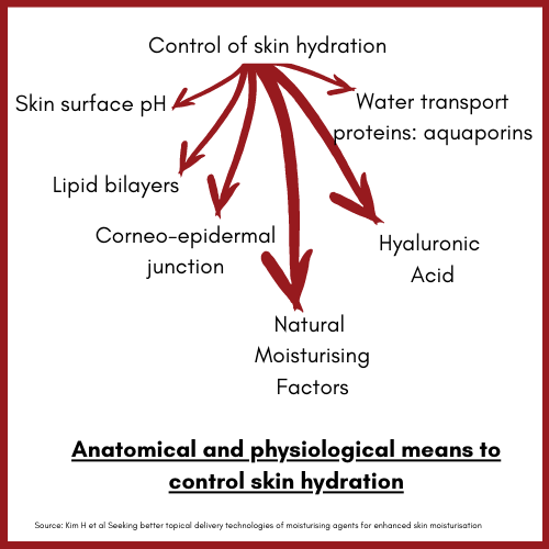
However, it is a mistake to belief that there are no other factors that contribute to skin hydration. I recently read a paper, which details the anatomical and physiological means by which the skin controls hydration. Its such an excellent paper, that I simply had to summarise it in today’s blog.
Kim et al identify 6 mechanism by which the skin’s anatomical ang physiological mechanisms control skin hydration:
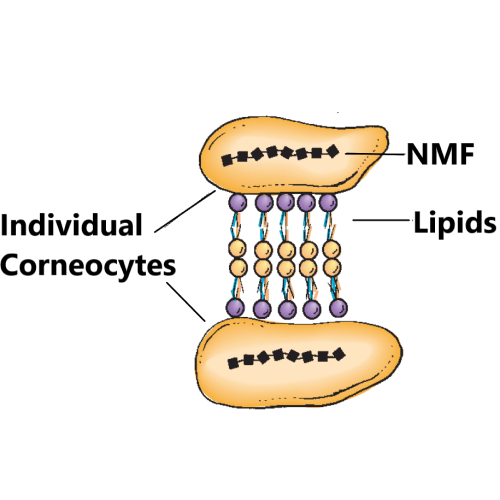
1. Natural Moisturizing Factors (NMF)
NMF components are found exclusively in the stratum corneum and are located in high concentrations within the corneocytes. The NMF consists primarily of amino acids (~40%) and their derivatives, including pyrrolidone carboxylic acid (PCA, ~12%), lactate (~12%), urea (~7%), and inorganic salts (~18%). ” (Source: Weber et al).
The corneocytes ae in essence “dead cells.” The NMFs within corneocytes are hygroscopic and act as an osmotic agent and draw water into the corneocytes (from environment and lower levels) to ensure that the appropriate level of water remains in the corneocytes
Source: Cosmetic Dermatology, by Dr Baumann (2nd edition)NMF come from (initially) insoluble protein called FILAGGRIN. Filaggrin is formed at the border of the Stratum Corneum / Stratum Granulosom and not before: if it did, then it would wreak havoc on lower layers of the skin.
This alignment of the corneocytes in the SC is what makes skin “soft” and “supple.” The NMF and lipids are at peace (or homeostasis) with the other elements around them. Both are essential for a proper functioning skin barrier. One of these crucial functions is reducing the amount of TEWL from the epidermis and keeping the skin hydrated.
If there were no Stratum Corneum, then we would dry out.
If the skin is dry (e.g., low humidity, age, medical condition such as diabetes etc.), then the corneocytes are not aligned tightly.
2. Hyaluronic Acid
Sakai et al (2000) were able to confirm the presence of HA in the Stratum Corneum. HA likely sits in the lamellar lipid matrix: it forms c.0.5% in weight of the NMFs. As a humectant, it attracts water from the environment and creates a voluminous structure.
HA in the dermis
HA is a component of the elastoviscous ECM in which collagen fibers, elastin and other structures are immersed. HA here has capacity to attract water in quantities equal to hundreds of times its weight. HA is therefore, a natural hydrating substance, responsible for tonicity of the skin and its reserves of moisture.
HA also facilitates transport of essential nutrients from the blood to skin cells.
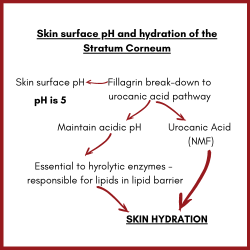
3. Skin surface pH
The skin surface pH is c5. Urocancic acid (UCA) which is a NMF is essential to maintaining the acidic environment of the skin.
The acidic environment is essential for hydrolytic enzymes essential to making of lipids to operate in. The lipid barrier reduces TEWL.
4. Lipid Bilayer and skin hydration
The lipid barrier is a permeability barrier that allows (e.g.) nutrients to pass through and excludes toxins but importantly it’s a waterproof barrier that protects TEWL. Major lipids in the SC that contribute to this permeability barrier are ceramides (50%), cholesterol (25%) and fatty acids (10%-20%).
The important parameter for maintaining skin moisturization is the 1:1:1 ratio of ceramides to cholesterol to fatty acids, in skin moisturisation products. Ceramides are riding a wave of popularity. They are important because of their weight and also their polar head groups provide adequate binding sites for water molecule.
5. Aquaporins (Water transporting proteins)
Kim et al write, “ Aquaporins are integral membrane proteins, that form water channels across biomembranes. Their principal function is to transport water, glycerol and other smaller solutes across membranes.
Aquaporins also facilitate the movement of water molecules within different layers of skin tissue and control hydration of deeper layers of the epidermis. The most abundant type of Aquaporin in the skin is AQP3 and its very relevant to skin hydration.
6. Corneo-epidermal junction
This is my representation of the coreno-epidermal junction – and its not very scientific, but it works for me.
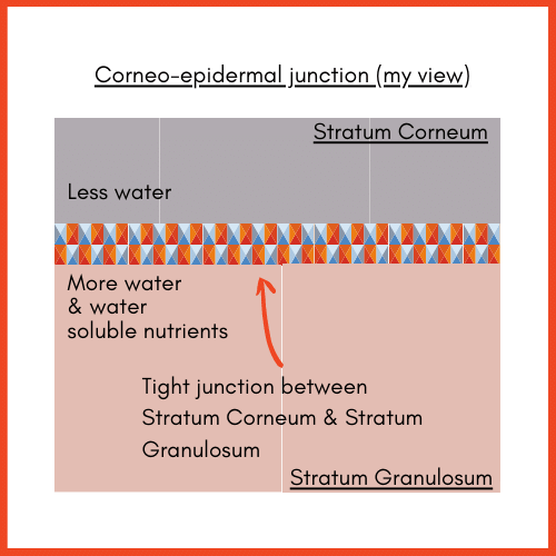
- The tight junction between the stratum corneum and the stratum granulosum maintains the diffusion of water from the lower viable dermis to the stratum corneum.
- The structure of the tight junction system consists of 40 transmembrane proteins (e.g. claudin, occludin) and JAM. The diffusion of water between spaces between the epidermal cells is principally regulated by claudins and occludins and JAMs. Also TJ ensure reduced TEWL by binding with barrier lipids.
Sources
Hyeongmin Kim, Jeong Tae Kim, Sonia Barua, Seung-Yup Yoo, Seong-Chul Hong, Kyung Bin Lee & Jaehwi Lee (2018) Seeking better topical delivery technologies of moisturizing agents for enhanced skin moisturization, Expert Opinion on Drug Delivery, 15:1, 17-31, DOI: 10.1080/17425247.2017.1306054